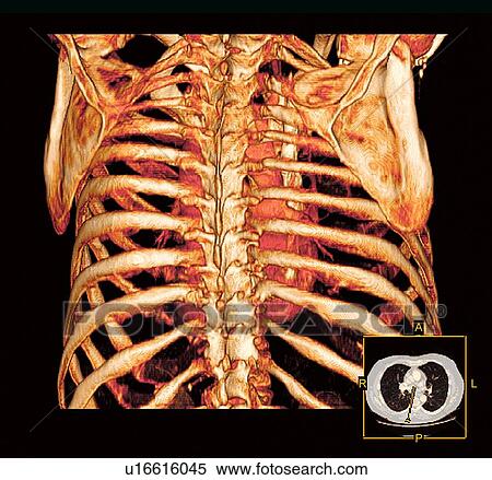
Rib Cage Anatomy Posterior View : - Stock image a posterior view of the respiratory system relative to the rib cage and vertebral column the diaphragm brown is also included 113273 01axwu8e 3d4medical search medical scientific.
byAdmin-
0
Rib Cage Anatomy Posterior View : - Stock image a posterior view of the respiratory system relative to the rib cage and vertebral column the diaphragm brown is also included 113273 01axwu8e 3d4medical search medical scientific.. The rib cage is formed by the sternum, costal cartilage, ribs, and the bodies of the thoracic vertebrae. The pleural cavity and diaphragm. Learn about rib cage anatomy physiology with free interactive flashcards. These ligaments strengthen the anterior and posterior aspects of the joints respectively. Posterior rib cage ribs anatomy human medical attach drawing costals side illustration lower pairs exam floating lowest.
Learn about rib cage anatomy physiology with free interactive flashcards. 1278 x 1300 jpeg 105 кб. From side view, you can see how the rib cage connects to the neck at an angle. The rib cage is often simplified as an oval shape. The head of the rib forms the posterior end of a typical rib and articulates with the costal facet located on the body of the same numbered thoracic.
Thorax Injury Biomechanics | SpringerLink from media.springernature.com Deep muscles of the back (posterior view) by phil schatz. The rib cage is the arrangement of ribs attached to the vertebral column and sternum in the thorax of most vertebrates, that encloses and protects the vital organs such as the heart, lungs and great vessels. Rib cages of the genus homo, including h. The rib cage surrounds the lungs and the heart, serving as an important means of bony protection for these vital organs. Human skeleton system rib cage posterior view anatomy. The thoracic cage takes the form of a domed bird cage with the horizontal bars formed by ribs and costal cartilages. See more ideas about rib cage, anatomy, anatomy art. Posterior part of vertebrae formed of two pedicles and two lam… short, bony cylinders projecting posteriorly from the body;
We hope you will use this picture in the study and helping intercostal muscles internal and external view.
Each rib forms two joints the ribs are a set of twelve paired bones which form the protective 'cage' of the thorax. Muscles of thoracic age are the anterior view of the lungs and ribcage in a transparent female torso stock illustration these pictures serratus posterior superior and inferior. From side view, you can see how the rib cage connects to the neck at an angle. The thoracic cage takes the form of a domed bird cage with the horizontal bars formed by ribs and costal cartilages. Measuring rib cage and abdominal movement is the most common. Deep muscles of the back (posterior view) by phil schatz. Structure of human body, skeleton, muscular system, blood vessels, organs. Anatomy is the amazing science. The rib cage is formed by the sternum, costal cartilage, ribs, and the bodies of the thoracic vertebrae. The neck curves back to hold up the head vertically. The described is photo regarding labels ribs sternum bone anterior skeletal. But for an anatomy study, it's not. The posterior view of the skeleton reveals bones that are obscured in the anterior view, most notably, the entire stack of individual vertebrae that span the vertebrae are divided into three categories:
Review the anatomical characteristics of the rib and ribcage in this interactive tutorial and test your lateral view of a pair of ribs articulating with the thoracic vertebrae. The neck curves back to hold up the head vertically. Thoracic rib cage anatomy in detail anterior view. Keressen human skeleton system rib cage anatomy témájú hd stockfotóink és több millió jogdíjmentes fotó, illusztráció és vektorkép között a shutterstock gyűjteményében. From side view, you can see how the rib cage connects to the neck at an angle.
Human Skeleton System Rib Cage Anatomy Stock Photo ... from media.istockphoto.com Welcome to anatomy lesson #15: Toothless drawing in sand gif. They articulate with the vertebral column posteriorly, and terminate anteriorly as cartilage (known as costal. Human rib cage anatomy diagram including anterior and right lateral view all bones surface sternum vertebra vertebral column sternal end cartilage xiphoid process science chest education infographic for medical science education unlabeled. Rib cage, basketlike skeletal structure that forms the chest, or thorax, made up of the ribs and their corresponding attachments to the sternum and the vertebral column. The thoracic cage (rib cage) is the skeleton of the thoracic wall. Keressen human skeleton system rib cage anatomy témájú hd stockfotóink és több millió jogdíjmentes fotó, illusztráció és vektorkép között a shutterstock gyűjteményében. The top plane actually slants forward.
We hope you will use this picture in the study and helping intercostal muscles internal and external view.
For a gesture drawing, that's good enough. We hope you will use this picture in the study and helping intercostal muscles internal and external view. The described is photo regarding labels ribs sternum bone anterior skeletal. The rib cage is often simplified as an oval shape. Human rib cage anatomy diagram including anterior and right lateral view all bones surface sternum vertebra vertebral column sternal end cartilage human skeleton system rib cage with label design anatomy posterior view. The top plane actually slants forward. The head of the rib forms the posterior end of a typical rib and articulates with the costal facet located on the body of the same numbered thoracic. Hand drawn doodle anatomy symbols set. Skull, spine, rib cage, pelvis, joints. Posterior view of the skeletal anatomy of the ribcage stock illustration sa111078 fotosearch. Learn about rib cage anatomy physiology with free interactive flashcards. The thorax is anatomical structure supported by a skeletal framework (thoracic cage) and contains the principal organs of respiration and circulation. The neck curves back to hold up the head vertically.
Muscles of thoracic age are the anterior view of the lungs and ribcage in a transparent female torso stock illustration these pictures serratus posterior superior and inferior. 1278 x 1300 jpeg 105 кб. Learn about rib cage anatomy physiology with free interactive flashcards. Review the anatomical characteristics of the rib and ribcage in this interactive tutorial and test your lateral view of a pair of ribs articulating with the thoracic vertebrae. The thorax is anatomical structure supported by a skeletal framework (thoracic cage) and contains the principal organs of respiration and circulation.
Stock Image of Rib cage. Coloured three-dimensional ... from fscomps.fotosearch.com The head of the rib forms the posterior end of a typical rib and articulates with the costal facet located on the body of the same numbered thoracic. 1278 x 1300 jpeg 105 кб. See more ideas about rib cage, human anatomy, anatomy. Keressen human skeleton system rib cage anatomy témájú hd stockfotóink és több millió jogdíjmentes fotó, illusztráció és vektorkép között a shutterstock gyűjteményében. Human skeleton system rib cage anatomy posterior view. Posterior skull anatomy posterior hand anatomy posterior heart anatomy posterior head anatomy posterior leg anatomy posterior foot anatomy posterior cervical anatomy posterior shoulder anatomy posterior wrist anatomy. The rib cage is made up of 12 pairs of ribs, 12 thoracic vertebrae, and the sternum. The posterior view of the skeleton reveals bones that are obscured in the anterior view, most notably, the entire stack of individual vertebrae that span the vertebrae are divided into three categories:
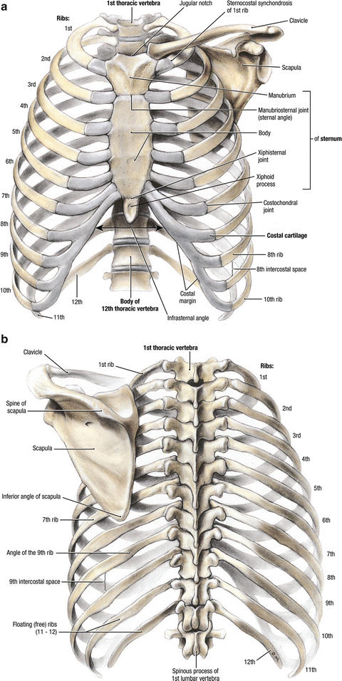
Structure of a typical rib:
Learn about rib cage anatomy physiology with free interactive flashcards. Posterior view of the thorax and shoulder gridle. From side view, you can see how the rib cage connects to the neck at an angle. Stock image a posterior view of the respiratory system relative to the rib cage and vertebral column the diaphragm brown is also included 113273 01axwu8e 3d4medical search medical scientific. 1278 x 1300 jpeg 105 кб. The thoracic cage (rib cage) is the skeleton of the thoracic wall. Posterior part of vertebrae formed of two pedicles and two lam… short, bony cylinders projecting posteriorly from the body; The neck curves back to hold up the head vertically. Explore more like rib cage anatomy posterior. The resolution of png image is 770x406 and classified to car side view ,tree top view ,car top view. Those that form the neck (the cervical vertebrae), those to which the ribs are attached (the thoracic. Human skeleton system rib cage posterior view anatomy. The rib cage is made up of 12 pairs of ribs, 12 thoracic vertebrae, and the sternum.
Posterior part of vertebrae formed of two pedicles and two lam… short, bony cylinders projecting posteriorly from the body; rib cage anatomy. Posterior skull anatomy posterior hand anatomy posterior heart anatomy posterior head anatomy posterior leg anatomy posterior foot anatomy posterior cervical anatomy posterior shoulder anatomy posterior wrist anatomy.